
To get an understanding of discarthrosis with or without spinal canal stenosis and your overall health, some tests are needed. The doctor will discuss with you which tests are required in your case.
EOS Imaging

During an EOS imaging, X-rays are used to simultaneously take an image of the spine and pelvis in the anterior/posterior and lateral directions. This is done to understand the influence of any arthrosis of the intervertebral disc that is present and any spinal canal stenosis on its position of the vertebrae among themselves in the standing position. The balance between the spine and pelvis is also examined.
Process:
- You take your position in the EOS device.
- You stand straight in a relaxed and natural manner while placing the fingertips on the collarbone.
- In the meanwhile, X-ray images are taken.
- While the images are being taken, you need to stand quietly and hold your breath for a moment.
- The total examination takes only 30 seconds.
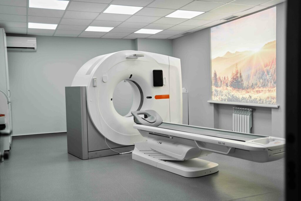
CT scan / SPECT-CT scan
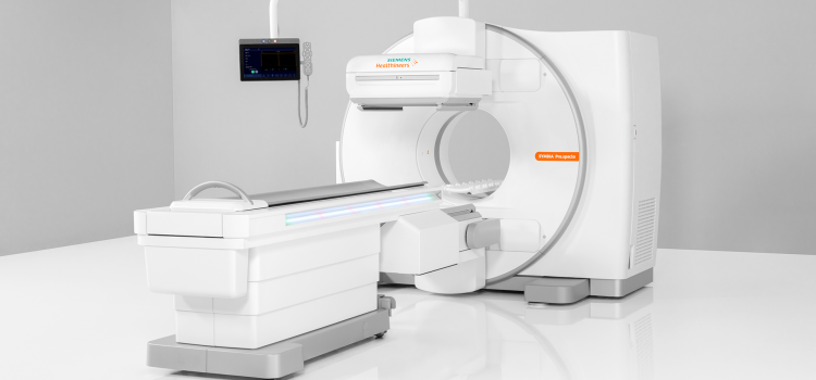
A CT scan uses X-rays and provides detailed cross sections of the spine. The cross sections are converted by the computer into images from different angles and into 3D images. When this is combined with a nuclear scan, it is called a SPECT-CT scan.
Process:
- Before the examination, a contrast medium may be injected through a vein in your arm.
- You will then take place on the examination table, usually on your back.
- The table is scrolled through a wide dome. Meanwhile, X-ray images are taken. While the images are being taken, you are required to lie quietly and hold your breath for a moment.
- The total examination takes an hour and a half where the CT scan itself takes about fifteen minutes.
MRI scan (Magnetic Resonance Imaging)
MRI scan provides information about the spinal cord, nerves, and muscles. MRI scan also makes cross sections of the spine. It uses short radio waves that generate signals in the body. Those signals are finally processed into images.
Process:
- You will lay down on the examination table.
- Before the examination, a contrast medium may be injected through a vein in your arm.
- The table slowly slides into a large tunnel. It is open on both sides and is well lit and ventilated.
- When the images are taken, you will hear tapping noises. So that the noise doesn’t bother you as much, you will be given headphones or earplugs.
- Stay as still as possible. Most often you will be asked to hold your breath for a moment.
- The medical team is outside the examination room but can always see you through a window and a camera.
- The examination takes twenty to thirty minutes, but in certain cases it may take an hour.
Considerations:
- Carefully read the guidelines on the appointment letter for your MRI examination.
- Complete the questionnaire on the appointment letter in advance and bring it with you to the examination. If anything is not clear, please contact your doctor to answer the questions together as correctly as possible.
- You do not need to be fasting for this examination.
- Please sign in at the hospital 45 minutes before your appointment.
- Do not wear metal objects (jewellery, piercings, hairpins).
- If you already have earplugs of your size at home, you can bring them with you to the examination.
- In case of (suspected) pregnancy and/or piercings or jewellery that you cannot remove, it is important to notify the nurse.
EMG (electromyography)
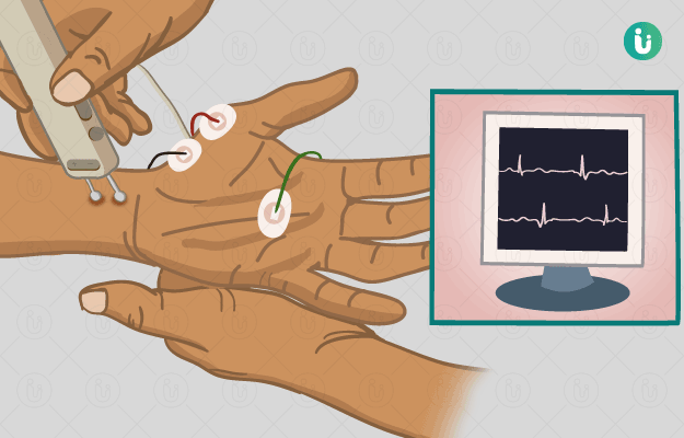
During an EMG, the function of the muscles and nerve pathways in the limbs is measured.
The test consists of two parts:
- The nerve test: You will receive electrical stimulation to the nerves, feeling small shocks. The response of the nerve is then accurately measured.
- The muscle test: A thin needle is inserted through the skin into the muscle, and you feel a prick. Depending on the muscle to be examined, you perform a certain movement for the doctor to see the action of the muscle on a computer screen.
Considerations:
- Make sure you are at the hospital 25 minutes before your appointment to register at least 10 minutes before your appointment.
- You do not need to be fasting.
- Clean your skin well with soap and water before the examination, but do not use skin creams.
- Please mention before the examination whether you take blood thinners (and which ones), cortisone-containing medication (e.g., puffers, skin creams, Medrol …), and whether you have received injections in the back or joints in the past two to three months.